Modern Microscopy
Linear confocal microscopy is a versatile tool for 3-dimensional image acquisition with sub-µm spatial resolution. Very often, however, linear scattering is not sensitive to the material properties or compositions of interest. Our research activities in the field of microscopy and optical analytics are therefore focused mainly on nonlinear techniques, which have the potential to provide new contrast mechanisms in many cases.
Second-harmonic imaging microscopy has been applied to obtain images of ferroelectric domain boundaries in periodically poled LiNbO3 structures. Performed in a confocal mode, with a modern fs-laser source and single photon detectors, this method provides a tomography of the domain boundaries by scanning the specimen with respect to the fixed laser focus.
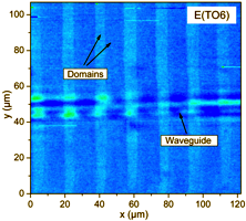
Raman imaging microscopy, also performed in a confocal mode, has been used for example to determine the strain distribution in pseudomorphic semiconductor nanostructures. Applied to ferroelectric domains, this method provides not only contrast to the domain boundaries, but also to the orientation of the individual domains. Because of its intrinsic sensitivity to the vibrational modes of molecular entities, Raman imaging is also a powerful tool to investigate the reaction kinetics in micro-channels under stationary flow conditions (funded by the Deutsche Forschungsgemeinschaft).


