Research & Facilities

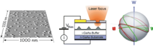
The research activities of the nanostructure optoelectronics group are focused on the physics and technology of semiconductor nanostructures and on the development and application of advanced optical analytics.

 Our work on semiconductor nanostructures is concentrated on the preparation and investigation of single quantum systems, their controlled manipulation and functionalization on the level of single electrons, excitons, photons or spins. This research field falls into the area of solid-state based quantum information technology, in which the coherent control of single quantum systems is of fundamental importance.

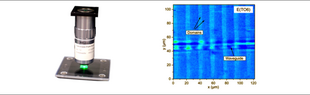
 In the field of optical analytics we are focused on fs-nonlinear confocal microscopy and Raman imaging. Applied to semiconductors, periodically poled ferroelectrics, and chemical reactions in micro-channels, those methods provide sub-µm spatial resolution and contrast mechanisms, which are inaccessible by linear optical microscopy.

Semiconductor quantum dots are artificial atoms contained in a host crystal. Their absorption and emission spectra appear as atomically sharp lines. By resonant optical excitation with a tunable laser system it is possible to generate or annihilate excitons (single electron-hole pairs) in the ground state of a quantum dot. With ps laserpulses it is possible to control excitons fast and efficiently in a defined way. Under those conditions, an exciton can be described as a quantum bit (qubit), which can be coherently manipulated in amplitude and phase.


For our experiments we use…

Linear  confocal  microscopy  is  a  versatile  tool  for  3-dimensional  image  acquisition  with sub-µm  spatial  resolution.  Very  often,  however,  linear  scattering  is  not  sensitive  to  the material  properties  or  compositions  of  interest.  Our  research  activities  in  the  field  of microscopy  and  optical  analytics  are  therefore  focused  mainly  on  nonlinear  techniques, which  have  the  potential  to  provide  new  contrast  mechanisms  in  many  cases.

Second-harmonic  imaging  microscopy  has  been  applied  to  obtain  images  of  ferroelectric  domain …